

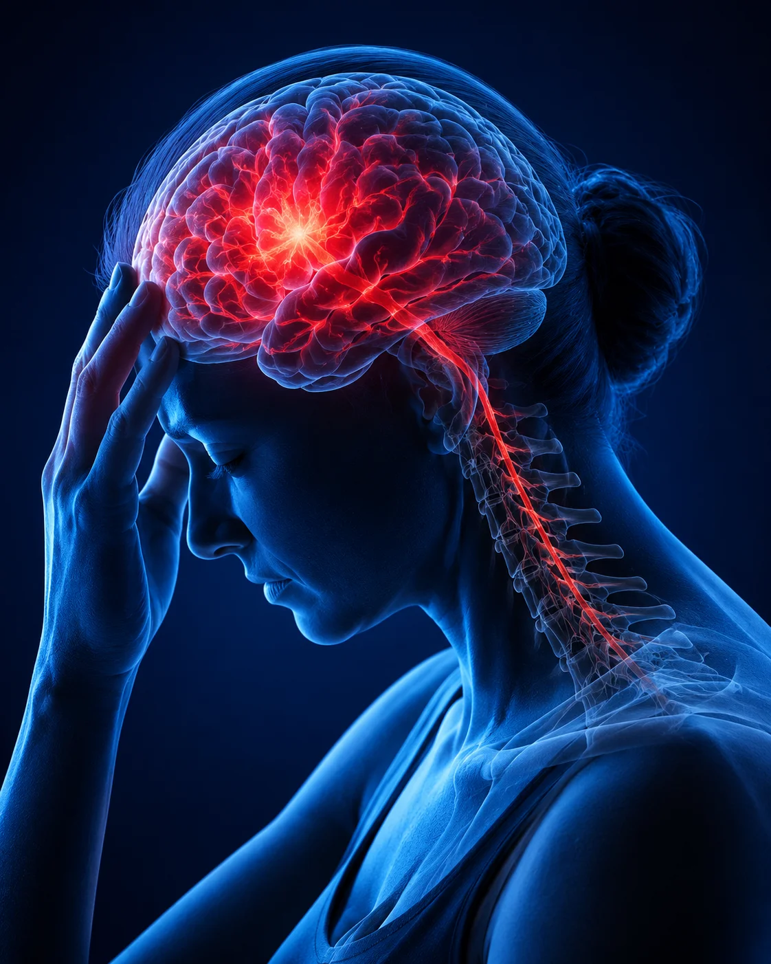
A CRH stress signal involved in the body’s response to stress may also have an important role in repairing the brain after injury, according to research led by scientists at the Max Planck Institute of Psychiatry in Germany. The study found that certain precursor cells surrounding an injured area of the brain temporarily produce corticotropin-releasing hormone (CRH), helping regulate the process by which damaged nerve insulation is restored.
The findings offer a new perspective on CRH, a neuropeptide widely associated with the body’s stress-response system. Rather than acting only as part of a stress pathway, CRH appears to participate directly in the local response to brain injury.
The research was published in Cell Reports on November 25, 2025, under the title Neuropeptide CRH prevents premature differentiation of OPCs following CNS injury and in early postnatal development. The paper was published by a research team involving scientists from the Max Planck Institute of Psychiatry and collaborating institutions.
CRH Stress Signal Appears to Guide Brain Repair
The study focused on oligodendrocyte progenitor cells (OPCs), precursor cells that can develop into oligodendrocytes. These specialized cells produce myelin, the insulating material that surrounds nerve fibers and helps them transmit electrical signals efficiently.
Following an acute brain injury, the researchers observed a previously underappreciated population of OPCs that begins producing CRH around the damaged area. The response occurs rapidly and is temporary.
According to the researchers, CRH expression can be detected within hours of an injury and largely subsides after roughly three days. The published research found CRH-positive OPCs around injury sites and linked their activity to the subsequent production of myelin-forming oligodendrocytes.
This timing appears to be important.
Instead of simply accelerating the production of new oligodendrocytes, the CRH system appears to help prevent precursor cells from differentiating too quickly. This allows the repair process to develop in a more controlled manner.
How OPCs Help Restore Damaged Nerve Fibers
Myelin is essential for normal communication within the nervous system. When it is damaged, nerve signals can become less efficient or disrupted.
OPCs serve as a pool of precursor cells that can respond to damage and generate new oligodendrocytes. These newly formed cells can then contribute to the restoration of myelin around affected nerve fibers.
The Max Planck research suggests that CRH produced by one population of OPCs communicates with another population carrying CRH receptor 1 (CRHR1).
This interaction appears to regulate the pace of oligodendrocyte development.
Researchers found that disrupting the CRH/CRHR1 system caused OPCs to generate oligodendrocytes more rapidly after acute injury. However, the faster response did not necessarily produce better long-term repair. Instead, the absence of normal CRHR1 signaling was associated with fewer mature oligodendrocytes surviving over time.
The findings indicate that speed is not the only factor in brain repair. The timing and maturation of repair cells also matter.
Why Timing Matters
The researchers’ results suggest that CRH acts as part of a biological control mechanism that prevents premature differentiation of OPCs.
In simple terms, the signal appears to tell precursor cells not to rush through the repair process.
When CRH/CRHR1 signaling was disrupted, the cells moved toward oligodendrocyte production more quickly, but the resulting repair population was less durable after acute injury. This suggests that a carefully controlled sequence of proliferation, differentiation and maturation may be necessary for effective restoration of myelin.
The Study Was Conducted in Laboratory Models
An important limitation is that these findings come from experimental research rather than clinical trials in people.
The study used mouse models and genetic approaches to investigate CRH-producing OPCs and CRHR1-expressing cells. The researchers examined how changing the CRH/CRHR1 system affected oligodendrocyte development, injury responses and myelin structure.
That means the findings should not yet be interpreted as evidence that increasing CRH in humans would improve recovery from a brain injury.
Further research will be needed to determine whether the same mechanism operates in people and whether it can eventually be targeted safely for therapeutic purposes.
Possible Connection to Stress and Psychiatric Disorders
The discovery may also be relevant beyond physical brain injuries.
CRH is already closely associated with the biological stress response. It is particularly well known for its role in regulating communication between the brain and the body’s hormonal stress system.
The researchers found that CRH signaling also influences oligodendrocyte development during early brain development. In their experiments, disruption of the CRH/CRHR1 pathway affected early postnatal oligodendrogenesis and produced changes in adult myelination.
This raises questions about whether changes in CRH signaling could contribute to some conditions involving stress, brain development or altered myelination.
The Max Planck researchers specifically pointed to possible relevance for stress-associated psychiatric disorders, including depression. However, this represents a potential area for future investigation rather than an established explanation for these conditions.
What the Findings Could Mean for Multiple Sclerosis
The study may also attract attention in research involving demyelinating diseases such as multiple sclerosis, in which the protective myelin coating around nerve fibers is damaged.
Because OPCs and oligodendrocytes are central to myelin production and repair, understanding how these cells respond to injury could eventually contribute to research into therapies designed to promote remyelination.
However, the study did not demonstrate a CRH-based treatment for multiple sclerosis, nor did it establish that manipulating CRH can repair myelin in patients.
The significance at this stage is more fundamental: researchers have identified a previously unrecognized signaling mechanism that influences how OPCs respond and mature after central nervous system injury.
A New View of the Brain’s Response to Injury
The research adds to growing evidence that the brain has complex internal mechanisms for responding to damage.
The discovery that OPCs themselves can temporarily produce CRH is particularly notable. Researchers had traditionally associated CRH more strongly with neurons and the broader stress-response system. The study identifies a distinct population of OPCs that can activate CRH following injury.
The response is also highly time-dependent. Researchers detected CRH-producing OPCs as early as 12 hours after injury, with the signal becoming more prominent during the first two days and declining substantially over subsequent days.
This suggests that the brain’s repair response is not simply a matter of switching regeneration on or off. Instead, different cellular signals may operate at specific stages to coordinate the repair process.
What Researchers Will Need to Study Next
The next step will be determining whether this mechanism can be translated into human neuroscience and medicine.
Scientists will need to establish whether CRH-producing OPCs and CRHR1-dependent signaling operate in the same way in the human brain. They will also need to determine whether manipulating this pathway could improve recovery without producing unwanted effects elsewhere in the body’s stress system.
Because CRH has important functions throughout the nervous and endocrine systems, any potential treatment targeting the pathway would likely require highly specific approaches.
For now, the study provides a new biological insight rather than an immediately available medical treatment.
Why the Research Matters
The significance of the findings lies in the connection between two areas that are often considered separately: stress signaling and neural repair.
The researchers have shown that CRH can be produced locally by precursor cells after brain injury and that its interaction with CRHR1 helps regulate the development and survival of oligodendrocytes.
The work therefore suggests that the brain may use a familiar stress-related signaling molecule as part of a carefully timed repair response.
The study does not mean that stress itself is beneficial for brain health. Instead, it reveals that a molecule involved in the stress system can have a different, locally controlled function inside injured brain tissue.
That distinction is important.
The findings could eventually help researchers better understand how the nervous system responds to injury, how myelin is maintained and repaired, and how disruptions in these processes might relate to neurological or psychiatric disorders.
For now, however, the evidence remains experimental, and more research is required before the discovery can be translated into treatments for patients.
Our audience means everything to us.
Have feedback or suggestions to make us better? We’re always listening—feel free to get in touch with us at editor@truthorahub.com









Leave a Comment